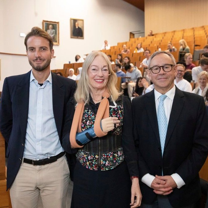
Peri-implant diseases, such as peri-implant mucositis and peri-implantitis, are significant complications affecting dental implants. However, the development of advanced biomaterials offers a promising solution. These materials have the potential to significantly reduce bacterial colonisation, promote tissue integration, and actively contribute to healing, instilling a sense of hope and optimism for the future of implantology.
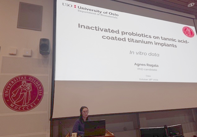
Our department was proud to present the innovative contributions of our researchers. Badra Hussain's work on chemical contamination on peri-implantitis, and Agnes Rogala's research on the use of inactivated probiotics on tannic acid-coated titanium implants, are unique and promising. These studies, which highlight the role of biomaterials in preventing biofilm formation and promoting healthy tissue responses, are crucial in reducing the incidence of peri-implant diseases.

Sadia Khan's presentation on using a chitosan brush to reduce peri-implantitis is a testament to the power of interdisciplinary collaboration. This research, conducted in partnership with our department, explores the effectiveness of chitosan-coated brushes in inhibiting bacterial growth and reducing inflammation around implants. The bioactive properties of chitosan show significant potential for limiting peri-implantitis progression, and Sadia Khan’s work highlights how such collaborations can drive forward new and effective solutions for implant-related challenges.
By exploring how surface modifications and bioactive coatings can enhance implant integration and reduce infection risks, our research team contributes to developing next-generation dental implants that can better withstand the challenges posed by peri-implant diseases. The seminar highlighted the work of international leaders in the field. It demonstrated the significant advancements being made by our researchers, further strengthening our position as a leading centre for biomaterials research. We thank all participants and presenters for making this seminar a success.
If you are interested in reading more about the research behind it, please find our latest published papers on how to combat biomaterial associated infections (BIA) with a brief summary below:
The research paper "Combining QCM-D with Live-Cell Imaging Reveals the Impact of Serum Proteins on the Dynamics of Fibroblast Adhesion on Tannic Acid-Functionalised Surfaces" investigates how tannic acid (TA) nanocoatings on titanium surfaces influence the adhesion of human gingival fibroblasts (hGFs). The study used quartz crystal microbalance with dissipation (QCM-D) monitoring and live-cell imaging to assess cell adhesion and behaviour in media with varying concentrations of fetal bovine serum (FBS). The results demonstrated that TA-coated surfaces enhanced the initial adhesion of fibroblasts in the absence or at low concentrations of FBS. In contrast, higher concentrations significantly inhibited cell adhesion and spreading. The findings indicate that TA coatings could provide a method for modulating cell adhesion on implant surfaces, potentially preventing excessive fibroblast attachment in biomedical applications. These insights suggest that controlling serum protein interactions can be crucial for successfully integrating implants and preventing fibrosis.
"Peri-Implant Health and the Knowing-Doing Gap—A Digital Survey on Procedures and Therapies" explores expert opinions on managing peri-implant health and treating peri-implant diseases. The survey revealed no consensus on the most effective treatment methods, with experts expressing concerns about current decontamination protocols and the overuse of antibiotics due to resistance risks. While mechanical cleaning methods like motor-driven brushes and ultrasonic devices were preferred, the lack of well-documented evidence and standardised protocols was highlighted. The study emphasises the need for further clinical research and standardised guidelines for managing peri-implant health.
"A Comparative Investigation of Chemical Decontamination Methods for In-Situ Cleaning of Dental Implant Surfaces" explores the effectiveness of various chemical agents in decontaminating titanium dental implant surfaces. The study evaluates the combination of Pluronic gel with hydrogen peroxide against other commercially available decontamination products. X-ray Photoelectron Spectroscopy (XPS) was used to analyse the chemical changes on the implant surfaces, and in vitro cytotoxicity tests assessed the safety of the chemical agents. The results revealed that NuBone®Clean demonstrated better efficacy in cleaning the implant surfaces, showing the highest titanium content and lowest nitrogen levels, indicating minimal residual contamination. None of the tested agents exhibited cytotoxic effects.
"Biofouling on Titanium Implants: A Novel Formulation of Poloxamer and Peroxide for In Situ Removal of Pellicle and Multi-Species Oral Biofilm" investigated the effectiveness of a new chemical decontamination solution, combining poloxamer gel with hydrogen peroxide in removing biofilms and preventing re-contamination on titanium implant surfaces. The study compares this formulation with other commonly used decontamination agents, such as EDTA, sodium hypochlorite (NaOCl), and chlorhexidine. The research focuses on two early stages of biofilm formation: the removal of the initially acquired pellicle and the prevention of its re-formation, as well as the reduction of static multi-species biofilms involving relevant bacteria like S. oralis, Fusobacterium nucleatum, and A. actinomycetemcomitans. The study demonstrates that while no single agent achieved complete biofilm removal, the combination of poloxamer and hydrogen peroxide showed a synergistic effect. It effectively reduced organic residues, enhanced surface hydrophilicity, and lowered re-contamination/reformation of pellicle rates compared to other tested agents.
The research paper "Anatomical Three-Dimensional Model with Peri-Implant Defect for In Vitro Assessment of Dental Implant Decontamination" introduces a new 3D-printed model designed to evaluate decontamination methods for dental implants. This anatomically realistic model mimics advanced peri-implant bone defects, allowing researchers to study the effectiveness of different mechanical and chemical decontamination tools in a controlled environment. The study used coloured occlusion spray to simulate biofilm on dental implants and applied various decontamination techniques, including an oscillating chitosan brush (OCB) and ultrasonic devices with polyetheretherketone tips (US-PEEK). The results demonstrated that the model enabled detailed analysis of biofilm removal and helped identify areas where decontamination could have been more effective. While no technique achieved complete decontamination, the model effectively differentiated the performance of each method and assessed changes in implant surface morphology post-treatment. This study concludes that the 3D-printed model is a valuable tool for testing and refining implant decontamination strategies. It offers an adaptable and reproducible way to study implant cleaning in vitro. Future research could further incorporate more complex defect shapes and dynamic biofilm simulations to validate its clinical relevance.


Photo credit: Marie Lindeman Johansen and Håvard J Haugen, OD/UiO